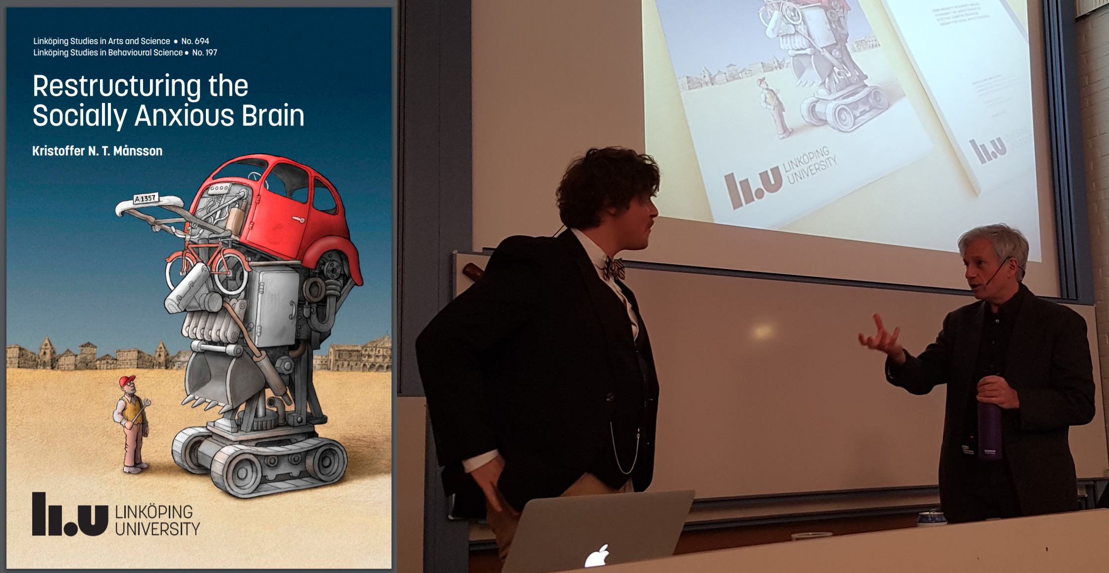
Today the psychologist and PhD student Kristoffer NT Månsson brilliantly defended his PhD thesis in Linköping University. The thesis was entitled “Restructuring the socially anxious brain: Using magnetic resonance imaging to advance our understanding of effective cognitive behaviour therapy for social anxiety disorder”. Philippe Goldin from University of California served as an excellent opponent. We, the supervisors (main: Gerhard Andersson, co-supervisors: Tomas Furmark, Per Carlbring, C-J. Boraxbekk) are very proud today.
[lightbox link=”http://www.carlbring.se/wp/wp-content/uploads/2016/12/IMG_20161215_162208.jpg” thumb=”http://www.carlbring.se/wp/wp-content/uploads/2016/12/IMG_20161215_162208-1024×766.jpg” width=”1024″ align=”center” title=”” frame=”true” icon=”image” caption=”From Left to Right: Tomas Furmark, Per Carlbring, Andreas Olsson (grading committee), Predrag Petrovic (grading committee), the NEW DR. Kristoffer NT Månsson, Gerhard Andersson, Philippe Goldin (opponent), C-J. Boraxbekk samt Mary Rudner (grading committee).”]
Here is the abstract from Kristoffer’s thesis (you can download the full thesis here):
Social anxiety disorder (SAD) is a common psychiatric disorder associated with considerable suffering. Cognitive behaviour therapy (CBT) has been shown to be effective but a significant proportion does not respond or relapses, stressing the need of augmenting treatment. Using neuroimaging could elucidate the psychological and neurobiological interaction and may help to improve current therapeutics. To address this issue, functional and structural magnetic resonance imaging (MRI) were repeatedly conducted on individuals with SAD randomised to receive CBT or an active control condition. MRI was performed pre-, and post-treatment, as well as at one-year follow-up. Matched healthy controls were also scanned to be able to evaluate disorder-specific neural responsivity and structural morphology. This thesis aimed at answering three major questions. I) Does the brain’s fear circuitry (e.g., the amygdala) change, with regard to neural response and structural morphology, immediately after CBT? II) Are the immediate changes in the brain still present at long-term follow-up? III) Can neural responsivity in the fear circuitry predict long-term treatment outcome at the level of the individual? Thus, different analytic methods were performed. Firstly, multimodal neuroimaging addressed questions on concomitant changes in neural response and grey matter volume. Secondly, two different experimental functional MRI tasks captured both neural response to emotional faces and self-referential criticism. Thirdly, support vector machine learning (SVM) was used to evaluate neural predictors at the level of the individual.
Amygdala responsivity to self-referential criticism was found to be elevated in individuals with SAD, as compared to matched healthy controls, and the neural response was attenuated after effective CBT. In individuals with SAD, amygdala grey matter volume was positively correlated with symptoms of anticipatory speech anxiety, and CBT-induced symptom reduction was associated with decreased grey matter volume of the amygdala. Also, CBT-induced reduction of amygdala grey matter volume was evident both at short- and long-term follow-up. In contrast, the amygdala neural response was weakened immediately after treatment, but not at one-year follow-up. In extension to treatment effects on the brain, pre-treatment connectivity between the amygdala and the dorsal anterior cingulate cortex (dACC) was stronger in long-term CBT non-responders, as compared to long-term CBT responders. Importantly, by use of an SVM algorithm, pre-treatment neural response to self-referential criticism in the dACC accurately predicted (>90%) the clinical response to CBT.
In conclusion, modifying the amygdala is a likely mechanism of action in CBT, underlying the anxiolytic effects of this treatment, and the brain’s neural activity during self-referential criticism may be an accurate and clinically relevant predictor of the long-term response to CBT. Along these lines, neuroimaging is a vital tool in clinical psychiatry that could potentially improve clinical decision-making based on an individual’s neural characteristics.
